
ARTIGO DE PESQUISA
Gel cream loaded with Lafoensia pacari phenolics-containing extract with antioxidant and photoprotective activities
https://doi.org/10.32712/2446-4775.2024.1228
- Caixêta, Eliane de Vasconcelos1
 https://orcid.org/0009-0001-0498-2713
- Diniz, Danielle Guimarães Almeida2
https://orcid.org/0000-0003-1867-3521
- Torres, Ieda Maria Sapateiro3
https://orcid.org/0000-0001-8407-627X
- Campos, Laila Portil Garcino1
https://orcid.org/0009-0001-7653-7176
- Machado, Rúbia Darc1
https://orcid.org/0000-0002-1811-8408
- Andréo, Bruna Galdorfini Chiari4
https://orcid.org/0000-0001-7959-0207
- Isaac, Vera Lucia Borges5
https://orcid.org/0000-0001-7402-920X
- Bara, Maria Teresa Freitas1*
https://orcid.org/0000-0003-4942-8721
- 1Federal University of Goiás (UFG), School of Pharmacy, Natural Products Research Lab (LPPN)/Bioproducts R&D&I Lab. Rua 240, corner of 5th Avenue, s/nº, University Sector. Zip Code 74.605-170, Goiânia, GO, Brazil.
- 2Federal University of Goiás (UFG), School of Pharmacy, Pharmaceutical Technology Lab. Alameda Flamboyant, Samambaia Technology Park, FarmaTec, University Campus, Zip Code 74690-310, Goiânia, GO, Brazil.
- 3Federal University of Goiás (UFG), School of Pharmacy, Laboratory of Quality Control of Medications, Rua 240, corner of 5th Avenue, s/nº, University Sector, Zip Code 74605-170, Goiânia, GO, Brazil.
- 4University of Araraquara (UNIARA). Rua Carlos Gomes, 1338, Centro, Zip Code 14801-340, Araraquara, SP, Brazil.
- 5São Paulo State University, Faculty of Pharmaceutical Sciences, Araraquara-Jaú Highway, km 1, University Campus, ZIP Code: 14800-850, Araraquara-SP, Brazil.
- *Correspondence:
- mtbara@gmail.com
Abstract
Lafoensia pacari`s leaves have phenolic compounds which may lead to photoprotective and antioxidant properties. The aim of this study was to investigate these activities in the L. pacari phenolics-containing extract (LPE) and to obtain gel cream formulations containing LPE and that have photochemoprotective activity. Phenolic compounds and antioxidant activity were determined. Polyacrylamide & C13-14 Isoparaffin & Laureth-7 (Focus gel® 305)formulation, it added synthetic sunscreen Ethylhexyl methoxycinnamate(Eusolex 2292®) or with both Eusolex 2292® and LPE were prepared. The in vitro sun protection factor (SPF) of these formulations was determined by diffuse reflectance spectroscopy. Preliminary stability of gel creams was evaluated. Total phenols and flavonoids content were 3.31% and 0.315%, respectively. LPE showed antioxidant activity by DPPH, with an EC50 of 5.3 µg. mL-1 and by FRAP methods, in which 1 g of the extract reduces 323.62 µM/L of ferrous sulfate. LPE showed an additive effect on the value of SPF when associated with Ethylhexyl methoxycinnamate,an increase of 22% in FPS of the formulation (F3). The data obtained allow us to suggest a trend towards the photoquimoprotective effect of the L. pacari phenolics-containing extract in the studied conditions.
- Keywords:
- Herbal medicinal.
- Sun protection factor.
- Additive effect on sun protection.
- Photochemoprotection.
Introduction
Polyphenols show important antioxidant[1-3] and photoprotective[4-6] activities.
Since the harmful effects of solar radiation can cause increased levels of reactive oxygen species (ROS) and reduced effectiveness of physiological antioxidant systems[5], the use of natural phenolics in topical formulation may be a strategy to be investigated, targeting useful anti-aging products for the prevention of skin aging[7].
Phytocompounds such as flavonoids, tannins, and others phenolics can act synergistically and influence the biological functions of the skin[8,9]. In addition, when these phenolic compounds are added to a formulation that contains chemical or physical sunscreens, they can, in combination, expand the sun protection factor, helping to make sunscreens more effective[4,10] and also enabling the use of low concentrations of synthetic sunscreen agents to prevent harmful effects of UV radiation[4].
A natural source of phenolic compounds is Lafoensia pacari A. St.-Hil. (Lythraceae), a tree-sized plant of Brazilian cerrado, which has antioxidant activity[11,12] and photoprotective potential[13]. The major chemical constituents reported are kaempferol and quercetin glycosides[14], catechin and ellagic acid derivatives[15], ellagitannins as punicalagin, punicalin and pedunculagin[14-16].
This study aimed to obtain a photoprotective and antioxidant formulation containing Lafoensia pacari phenolics-containing extract (LPE) and evaluate its ability to potentiate the action of a synthetic chemical sunscreen commonly used in cosmetics for this purpose.
Materials and Methods
Plant material and preparation of LPE
Lafoensia pacari leaves were collected in Caldazinha-GO (16º39'54.5''S, 49º00'03.9'' W, 1100 m altitude). A voucher specimen (UFG-47581) was deposited at the UFG Herbarium.
The leaves were dried in a forced-air circulation oven at 30°C, then ground in a knife mill and stored in a freezer. To obtain the L. pacari phenolics-containg extract of (LPE), 30g dry leaves were weighed, then 200 ml 70% alcohol were added and subjected to an ultrasonic bath for 15 minutes and finally was filtered[17].
Chemical characterization of LPE
The chromatographic profile by TLC was used. 10 µL of the extract were applied in silica gel plates with fluorescence indicator (Macherey-Nagel). The compounds were eluted using ethyl acetate: acetic acid: formic acid: distilled water in the ratio 100: 11: 11: 26 as mobile phase. The plate was revealed with NP (Sigma) 1% in methanol and placed under a 365 nm UV lamp. The presence of flavonoids (yellow bands) and phenolics acids (bluish white bands) were possible due to use of NP as revealer[18].
The total polyphenol content was determined by the Hagerman and Butler method[19]. The total flavonoid content was also determined[20].
To find the UV absorption spectrum of LPE, the maximum absorption wavelength was scanned in Waters® high performance liquid chromatography apparatus (HPLC), with e2695 separation module, a Waters® 2998 ultraviolet diode array detector equipped with Empower2® Build 2154 software. A Zorbax C18 column (250 x 4.6 mm, 5µm) was used, protected by a Phenomenex Security Guard C18 and maintained at 25°C. The reading was taken at 210 nm and the mobile phase flow (1.0 mL/min) was methanol: water (15:85)[21].
Antioxidant activity of LPE
The antioxidant activity was characterized by DPPH and FRAP methods. The DPPH method assay was performed[22]. Briefly, 100 µL of LPE (20 to100 µg/mL), positive control (ascorbic acid) and negative control (methanol) were mixed with 3.9 mL of 0.1 mM DPPH methanol solution. After 45 min of incubation at room temperature, the DPPH free radical reduction was measured at 515 nm. The radical scavenging activity was calculated using the linear regression analysis. The antioxidant properties were expressed as % DPPH inhibition. The extract concentration providing 50% of free radical scavenging activity (EC50) was calculated from the graph of the radical scavenging activity percentage against extract concentration.
Antioxidant activity based on iron reduction using the FRAP assay was also performed[23]. Briefly,90 µL of LPE (20 to 50 µM) were added to 2.7 ml of FRAP reagent (25 mL acetate buffer (300 mM) (pH 3.6), 2.5 mL TPTZ (Tri-pyridyl-triazine) solution (10 mM), and 2.5 mL FeCl3. 6H2O (20 mM) in aqueous solution). After 30 min at 37°C, the absorbance was measured at 595 nm, using the FRAP reagent as a blank. An aqueous solution of FeSO4 + H2O (0-2000 μmol/L) was used to prepare the calibration curve. The antioxidant capacity was expressed in μmols equivalent to Fe+2/L.
Formulations studied
The cosmetic base used was Focus gel 305® (Polyacrylamide & C13-14 Isoparaffin & Laureth-7; Lot: A15 /1595), one auto-emulsifiable base for the formulation of O/W emulsions.
Formulations (F1, F2 and F3) (TABLE 1) were prepared according to the traditional method to obtain a gel cream. The LPE and ethylhexyl methoxycinnamate(Eusolex 2292®: synthetic sunscreen tested)were incorporated in the gel cream until there was perfect homogeneity.
| Formulations | F1 | F2 | F3 |
|---|---|---|---|
| Polyacrylamide & C13-14 Isoparaffin & Laureth-7 | 5 | 5 | 5 |
| 4-hydroxybenzoic acid methyl ester | 0.15 | 0.15 | 0.15 |
| Ethylenediaminetetraacetic acid (EDTA) | 0.1 | 0.1 | 0.1 |
| Ethylhexyl methoxycinnamate | - | 6 | 6 |
| LPE | - | - | 5 |
| Distilled water (added to achieve 100%) | 100 | 100 | 100 |
In vitro sun protection factor (SPF)
The SPF for formulations containing synthetic sunscreen and the association of synthetic sunscreen and LPE was assessed by the Optometric SPF-290S Analyzer. The samples were prepared by spreading 110 mg of each formulation over a Transpore® tape (70.7 × 70.7 mm) to obtain a film of 2 g/cm2, as specified by the U.S. Food and Drug Administration. Each sample was exposed to a xenon arc solar simulator, and the Analyzer performed scans in 12 different spots on the Transpore® tape substrate. The scan was carried out, by measuring the transmittance, every 2 nm, in a wavelength range from 290 to 400 nm[24].
Preliminary stability and microbiological tests of formulations
Three batches of each formulation were prepared, stored in an inert plastic bottle.The product was kept at 25°C for 24 h after its preparation (To) and then analyzed.
For the centrifugation stability test, performed on 24 h-old formulations (F1 e F3), aliquots of approximately 10 g of the sample were weighed and packed in graduated Falcon-type tube and centrifugation (Sigma 3-18K) at 3000 rpm for 30 min at room temperature. The appearance, homogeneity and organoleptic characteristics were evaluated by macroscopic analyses.
For the thermal stress tests, formulations were submitted to a heated thermostatic bath (Fisatom) set for a temperature range of 40 to 80°C, with a temperature increase at intervals of 5°C, and held at each temperature for 30 min. The organoleptic characteristics, pH value (Gehaka PG 1800) and electrical conductivity (Tecnal TEC-4MP) measures were obtained to evaluate the formulations before and at the end at 80°C, after the natural cooling of the samples at room temperature (25 ± 2°C).
For freeze-defrost cycles (F/DF), samples were subjected to 4 ± 2°C/24 h (Electrolux, Air Flow System DC) and then 45 ± 2°C/24 h and 75% ± 5% RH (Nova Etica), thus completing 12 cycles. The organoleptic characteristics, pH value determination, and electrical conductivity measures were evaluated before (T0) and after preliminary stability tests. The pH value (Gehaka PG 1800) and electrical conductivity measures (Tecnal TEC-4MP) were determined at a temperature of 25 ± 2 °C. Viscosities were determined by Brookfield viscometer at a speed of 60 rpm coupled to the R7 spindle[25,26].
All analyses were conducted in triplicateand expressed as mean ± SD. Statistical analysis, ANOVA and Tukey tests were carried out when necessary, using GraphPad Prism 5.3.
The counting of the total number of mesophilic microorganisms was performed in F3 formulation before and after the freeze-defrost cycles using casein-soya agar at 32.5°C ± 2.5°C and Sabouraud Dextrose agar at 22.5°C ± 2.5°C, and incubated, respectively for 5 and 7 days. Escherichia coli, Salmonella sp., Pseudomonas aeruginosa, bile tolerant gram-negative bacteria and Staphylococcus aureus searches were performed[27].
Results and Discussion
Obtaining and chemical characterization of LPE
To obtain LPE, 70% ethanol was used as a vehicle extractor because this proportion of water and ethanol favors polyphenol extraction[17].
In TLC analysis, 7 bands could be seen indicating the presence of flavonoids (yellow bands) with a retention factor of 0.43, 0.73 and 0.90, and phenolic acids (bluish white bands) with a retention factor of 0.16, 0.64, 0.70 and 0.95. The aim of this TLC was to identify the class of secondary metabolites present in LPE and according to the revealer used (NP), bands of phenolic acids and flavonoids develop specific staining[18], as previously described.
Phenolic acids and flavonoids are important secondary metabolites that act as UV blockers. These compounds can prevent penetration of radiation into the skin, resulting in the reduction of inflammation, oxidative stress, and DNA damaging effects[4]. Thus they could enhance the final protection of the product and/or neutralize the free radicals produced in the skin after sun exposure[5].
After identifying these compounds, total phenols and flavonoids in LPE were quantified yielding 3.31% and 0.315%, respectively. To ensure the protection efficacy of sun-care products, the quantity of active constituents in natural extracts, compatibility, concentration, and stability is a concern[4].
Another chemical aspect investigated was the absorption spectrum of LPE, which showed maximum absorption at wavelength 258 nm, which corresponds to the UVC range, but LPE also showed an absorption range in the UVB region (280 a 315 nm). Compounds with aromatic rings can absorb UV rays, especially UVA and UVB at a wavelength range of 200-400 nm[4,5].
Antioxidant activity
For DPPH method, an analytical curve was achieved (y= -0.1516x + 0.6288), presenting a linear correlation coefficient (r=0.9917) and allowed determinationof EC50 value. LPE presented great antioxidant activity, with EC50 of 5.3 µg.mL-1. The EC50 of ascorbic acid was 2.1 µg.mL-1. L. pacari extract showed EC50 value of 8.5 μg/mL[14].
The antioxidant activity of LPE by the FRAP method was also performed. The analytical curve of the ferrous sulfate was obtained (y=0.0007x - 0.026) presenting a linear correlation coefficient (r=0.9988) and was used to calculate the antioxidant activity. The result shows that1g of the extract reduces 323.62 µM/L of ferrous sulfate. Sucupira et al. [23] demonstrated that acerola fruit reduces by a 148 µM/L of ferrous sulfate. The LPE reduces about 50% more ferrous sulfate than acerola fruit, thereby corroborating its antioxidant power.
The antioxidant capacity observed in this study can be attributed to the content of phenolic compounds.
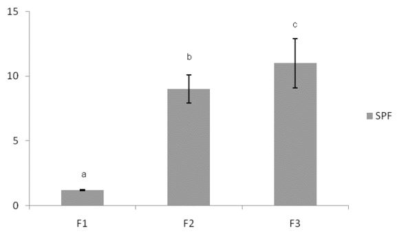
In vitro sun protection factor
The SPF values were estimated by the spectrophotometric method in the UV region (290-400 nm) using a Diffuse Reflectance Spectroscopy (FIGURE 1). The SPF for synthetic sunscreen used was about 9, while the SPF for synthetic sunscreen associated to LPE resulted in an improvement of 22%. This result showed an additive effect between the synthetic sunscreen ethylhexyl methoxycinnamate and LPE. The use of natural compounds in combination with synthetic agents may provide an effective strategy for preventing harmful effects of UV radiation[5].
These results presented showed that LPE has the potential to have an additive effect on SPF when associated with synthetic sunscreen tested in formulations. We suggest that in addition to reducing the skin exposure to ionizing radiation, LPE antioxidant activity may also contribute to scavenge ROS, originated from the skin's cell metabolism or even those created by radiation that were not blocked; thus, showing a photochemoprotective effect. One advantage pointed out in relation to the additive effect on SPF is the possibility of reducing the concentration of the synthetic sunscreen, which has been desired[4,28], and an additional study with different concentrations of LPE in the formulation may contribute to this context.
Preliminary stability and microbiological tests
We used Focus gel 305® (Polyacrylamide & C13-14 Isoparaffin & Laureth-7) that is an auto-emulsifiable base for the formulation of O/W emulsions highly stable. Besides it has the advantages of being water soluble, easy incorporation, promotes non-oily gel-cream texture, cold handling, does not require mechanical agitation for small amounts, it presents wide range of pH stability, 2 to 11 (manufacturer information).
The F1 formulation was shiny white, with characteristic odor, while the F3 presented with characteristic odor and was a bright cloudy yellow, because of the extract. These macroscopic characteristics observed at zero were maintained after the thermal stress and freeze-defrost cycles. A slight modification of the organoleptic characteristics was observed at temperatures of 75°C, but no phase separation was observed during the tests.
The results obtained after freeze-defrost cycles, in which the formulations showed significant differences in terms of conductivity and viscosity before and after the said cycle (p <0.001), and the results of pH determination of the formulations were not affected by temperature changes (p> 0.05) during these preliminary stability tests (FIGURE 2). The conditions under which the study is conducted are not intended to estimate a useful life of the product, but assist in the screening of formulations[26]. Modifications in conductivity and viscosity values allow to detect cremation, sedimentation or phase inversion[29]. Thus, we may suggest that the observed changes in conductivity and viscosity values may be due to phase inversion. It indicates that the proposed formulation will need some adjustments for future studies, in addition to accelerated stability and long-term stability tests being carried out. We highlight here that our objective in this initial study was to investigate the photoprotective effect of LPE in gel cream formulation.

The microbiological analysis of the mesophilic microorganism count and search for pathogens in F3, before and after the 12 days of freeze-defrost cycles showed no growth for Staphylococcus aureus, Pseudomonas aeruginosa, Escherichia coli, Salmonella sp. or bile tolerant gram-negative bacteria. For total yeast, molds and bacteria counts, growth was seen to be lower than 10 UFC/g. These results are within recommended Brazilian parameters[26,27].
Conclusion
Lafoensia pacari phenolics-containing extract (LPE) showed potential to perform a photochemoprotective activity, since it presented photoprotective and antioxidant effects. We highlight its ability to improve the SPF of the synthetic sunscreen Ethylhexyl methoxycinnamatein formulation F3, under the experimental conditions used.
Funding
FINEP, CAPES and CNPq.
Conflict of Interest
The authors declare that they have no conflicts of interest.
Acknowledgements
Authors thank FINEP, CAPES and CNPq for its financial support for this study.
Contributions
Study design: DGAD; MTFB
Data curation: EVC; DGAD; MTFB
Data collection: EVC; LPGC; RDM; BGCA; VLBI
Data analysis: EVC; VLBI; DGAD; IMST; MTFB
Writing of the original manuscript: EVC; DGAD; MTFB
Writing review and editing: DGAD; IMST; MTFB.
References
1. Alonso C, Rubio L, Touriño S, Martí M, Barba C, Fernández-Campos F, et al. Antioxidative effects and percutaneous absorption of five polyphenols. Free Radic Biol Med. 2014; 75: 149-155. ISSN: 0891-5849. [https://doi.org/10.1016/j.freeradbiomed.2014.07.014].
2. Aloqbi A, Omar U, Yousr M, Grace M, Lila MA, Howell N. Antioxidant activity of pomegranate juice and punicalagin. Nat Sci. 2016; 8(6): 235-246. ISSN: 2698-6248. [https://doi.org/10.4236/ns.2016.86028].
3. Moilanen J, Karonen M, Tähtinen P, Jacquet R, Quideau S, Salminen JP. Biological activity of ellagitannins: effects as antioxidants, pro-oxidants and metal chelators. Phytochemistry. 2016; 125: 65-72. ISSN: 0031-9422.[https://doi.org/10.1016/j.phytochem.2016.02.008].
4. Saewan N, Jimtaisong A. Natural products as photoprotection. J Cosmet Dermatol. 2015; 14(1): 47-63.ISSN: 1473-2165.[https://doi.org/10.1111/jocd.12123].
5. Cefali LC, Ataide JA, Moriel P, Foglio MA, Mazzola PG. Plant-based active photoprotectants for sunscreens. Int J Cosmet Sci.2016; 38(4): 346-353. ISSN: 1468-2494.[https://doi.org/10.1111/ics.12316].
6. Carvalho WLR, Moreira LC, Valadares MC, Diniz DGA, Bara MTF. Pterodon emarginatus hydroalcoholic extract: antioxidant and photoprotective activities, noncytotoxic effect, and perspective of obtaining formulations with photochemoprotective activity.Pharmacogn Mag. 2019; 15(64): 176-182. ISSN: 0976-4062 [https://doi.org/10.4103/pm.pm_580_18].
7. Binic I, Lazarevic V, Ljubnovic M, Mojsa J, Sokolovic D. Skin ageing: natural weapons and strategies.Evid Based Compl Alt Med. 2013; 2013: 1-10. ISSN: 1741-4288. [https://doi.org/10.1155/2013/827248].
8. Fonseca-Santos B, Correa MA, Chorilli M. Sustainability, natural and organic cosmetics: consumer, products, efficacy, toxicological and regulatory considerations. Braz J Pharm Sci. 2015; 51(1): 17-26. ISSN: 2175-9790. [https://doi.org/10.1590/S1984-82502015000100002].
9. Chiorcea-Paquim AM, Enache TA, De Souza EG., Oliveira-Brett AM. Natural phenolic antioxidants electrochemistry: towards a new food science methodology. Compr Rev Food Sci Food Saf. 2010; 19(4): 1680-1726. ISSN: 1541-4337. [https://doi.org/10.1111/1541-4337.12566].
10. Daher CC, Fontes IS, Rodrigues RO, Damasceno GAB, Soares DS, Aragão CFS, et al. Development of O/W emulsions containing Euterpe oleracea extract and evaluation of photoprotective efficacy. Braz J Pharm Sci. 2014; 50(3): 639-652. ISSN: 2175-9790. [https://doi.org/10.1590/S1984-82502014000300024].
11. Solon S, Lopes L, Sousa JRPT, Schmeda-Hirschmann G. Free radical scavenging activity of Lafoensia pacari.J Ethnopharmacol. 2000;72(1-2): 173-178. ISSN: 0378-8741. [https://doi.org/10.1016/S0378-8741(00)00233-6].
12. Garcia SAS, Rocha PBR, Souza BS, Paz ATS, Negris ALC, Marreto RN, et al. Enhanced skin permeation of punicalagin after topical application of pluronic micelles or vesicles loaded with Lafoensia pacari extract. Pl Med. 2022; 88(6):479-488.ISSN: 0032-0943. [https://doi.org/10.1055/a-1464-1525].
13. Violante IMP, Souza IM, Venturini CL, Ramalho AFS, Santos RAN, Ferrari M. In vitro sunscreen activity evaluation of plants extracts from Mato Grosso cerrado.Braz J Pharmacogn. 2009; 19(2A): 452-457. ISSN: 0102-695X. [https://doi.org/10.1590/S0102-695X2009000300020].
14. Pereira LOM, Vilegas W, Tangerina MMP, Arunachalam K, Balogun SO, Orlandi-Mattos PE, et al. Lafoensia pacari A. St.-Hil.: Wound healing activity and mechanism of action of standardized hydroethanolic leaves extract. J Ethnopharmacol. 2018; 219: 337-350. ISSN: 0378-8741. [https://doi.org/10.1016/j.jep.2018.02.038].
15. Reichert CL, Silva DB, Carollo CA, Weffort-Santos AM, Santos CAM. Metabolic profiling and correlation analysis for the determination of killer compounds of proliferating and clonogenic HRT-18 colon cancer cells from Lafoensia pacari. J Ethnopharmacol. 2018; 224: 541-552. ISSN: 0378-8741. [https://doi.org/10.1016/j.jep.2018.06.021].
16. Carneiro CC, Santos SC, Lino RS, Bara MTF, Chaibub BA, Reis PRM, et al. Chemopreventive effect and angiogenic activity of punicalagin isolated from leaves of Lafoensia pacari A. St.-Hil. Toxicol Appl Pharmacol. 2016; 310: 1-8. ISSN: 1096-0333.[https://doi.org/10.1016/j.taap.2016.08.015].
17. Dutra RC, Leite MN, Barbosa NR. Quantification of phenolic constituents and antioxidant activity of Pterodon emarginatus Vogel seeds.Int J Mol Sci. 2008; 9(4): 606-614. ISSN: 1468-2494.[https://doi.org/10.3390/ijms9040606].
18. Wagner H, Bladt S. Plant Drug Analysis: A thin layer chromatography atlas. 2nd ed.Germany: Springer, 2001. 384 p. ISBN-10: 3540586768.
19. Mole SA, Waterman PG. A critical analysis techniques for measuring tannins in ecological studies. Oecologia. 1987; 72(1): 137-147. ISSN: 1432-1939.[https://doi.org/10.1007/BF00385058].
20. Rolim A, Maciel CPM, Kaneko TM, Consiglieri VO, Salgado-Santos IMN, Velasco MVR. Validation assay for total flavonoids, as rutin equivalents, from Trichilia catigua Adr. Juss. (Meliaceae) and Ptychopetalum olacoides Bentham (Olacacea) commercial extract. J AOAC Int. 2005; 88(4): 1015-1019. PMID: 16152916. ISSN: 1944-7922.[https://pubmed.ncbi.nlm.nih.gov/16152916/].
21. Oliveira LAR. Isolamento, quantificação e avaliação das atividades leishmanicida e tripanocida de furanoditerpenos do oleorresina de Pterodon spp. Vogel (Fabaceae). 119 p. Goiânia; 2014. Dissertação de Mestrado [Programa de Pós-Graduação em Ciências Farmacêuticas] - Universidade Federal de Goiás. Goiânia; 2014.
22. Lugato D, Simão M, Garcia R, Mansur E, Pacheco G. Determination of antioxidant activity and phenolic content of extracts from in vivo plants and in vitro materials of Passiflora alata Curtis. Plant Cell Tiss Organ Cult. 2014; 118(2): 339-346. ISSN: 1573-5044. [https://doi.org/10.1007/s11240-014-0486-4].
23. Sucupira NR, Silva AB, Pereira G, Costa JN. Methods for measuring antioxidant activity of fruits. UNOPAR Cient Cienc Biol Saúde. 2012; 14(4): 263-269. ISSN: 1517-2570.[https://doi.org/10.17921/2447-8938.2012v14n4p%25p].
24. Chiari-Andréo BG, Trovatti E, Pecoraro E, Correa MA, Cicarelli RMB, et al. Synergistic effect of green coffee oil and synthetic sunscreen for health care application. Ind Crops Prod. 2014; 52: 389-393.ISSN: 0926-6690.[https://doi.org/10.1016/j.indcrop.2013.11.011].
25. Ribeiro RCA, Barreto SMAG, Ostrosky EA, Rocha-Filho PA, Verissimo LM, Ferrari M. Production and characterization of cosmetic nanoemulsions containing Opuntia ficus-indica (L.) Mill extract as moisturizing agent.Molecules. 2015; 20(2): 2492-2509.ISSN: 1420-3049. [https://doi.org/10.3390/molecules20022492].
26. Brasil. Ministério da Saúde. Agência Nacional de Vigilância Sanitária. Guia de Estabilidade de Produtos Cosméticos. 1ª ed. Brasília: Anvisa, 2004. 52 p.
27. Brasil. Ministério da Saúde. Agência Nacional de Vigilância Sanitária. Brazilian Pharmacopeia, 5ª ed. Brasília: Anvisa. 2010. p.236-253. ISBN: 9788588233409.
28. Jansen R, Wang SQ, Burnett M, Osterwalder U, Lim HW. Photoprotection: part I. Photoprotection by naturally occurring, physical, and systemic agents. J Am Acad Dermatol. 2013; 69(6): 853-865. ISSN: 0190-9622. [https://doi.org/10.1016/j.jaad.2013.08.021].
29. Masmoudi H, Dréau YL, Piccerelle P, Kister J. The evaluation of cosmetic and pharmaceutical emulsions aging process using classical techniques and a new method: FTIR. Int J Pharm. 2005; 289(1-2): 117-131. ISSN: 0378-5173. [https://doi.org/10.1016/j.ijpharm.2004.10.020].