QUÍMICA
Bioguided isolation of an antiviral compound from Xylophragma myrianthum (Cham.) Sprague (Bignoniaceae Juss.)
Abstract
Xylophragma Sprague species (family Bignoniaceae Juss.) are climbing plants belonging to the tribe Bignonieae Juss. and some species have a wide spectrum of traditional medicinal uses including remedies for the treatment of infections. This paper reports the bioguided fractionation of an ethanol extract of X. myrianthum (Cham.) Sprague stems (EEXMS) for antiviral effects against Human herpesvirus 1 (HSV-1), Dengue virus 2 (DENV-2), murine Encephalomyocarditis virus (EMCV) and Vaccinia virus (VACV) that afforded XM-1 as an active compound. Spectroscopic analyses allowed the identification of XM-1 as arjunic acid whose occurrence in the Bignoniaceae and anti-DENV-2 activities are reported for the first time. X. myrianthum is revealed herein as a source of an antiviral compound and fractions.
- Key words:
- Triterpene.
- Arjunic acid.
- Dengue virus.
- MTT assay.
Resumo
Espécies do gênero Xylophragma Sprague são trepadeiras pertencentes à família Bignoniaceae Juss. (tribo Bignonieae Dumort.) e algumas tem um amplo espectro de usos medicinais, incluindo o tratamento de infec ções. No presente artigo relatamos o fracionamento do extrato etanólico de caules de Xylophragma myrianthum (Cham.) Sprague biomonitorado por testes de atividade contra Human herpesvirus 1 (HSV-1), Dengue virus 2 (DENV-2), Encephalomyocarditis virus murino (EMCV) e Vaccinia vírus (VACV), o que levou ao isolamento de uma substância ativa, XM-1. Análises espectroscópicas permitiram a identificação desta como sendo o triterpeno ácido arjúnico, cuja atividade anti-DENV-2 e ocorrência em Bignoniaceae são relatadas pela primeira vez. X. myrianthum revela-se, portanto, como fonte de uma substância e frações antivirais.
- Palavras Chave:
- Triterpeno.
- Ácido Arjúnico.
- Vírus Da Dengue.
- Ensaio do MTT.
Introduction
Xylophragma Sprague species are climbing plants belonging to the family Bignoniaceae Juss. (tribe Bignonieae Dumort.). Several representatives of the Bignoniaceae have a wide spectrum of traditional medicinal uses including remedies for the treatment of infections. The family Bignoniaceae comprises about 82 genera and 827 species that are distributed mainly in tropical regions around the world (Gentry, 1992; Lohmann, 2006). This botanical family consists of trees, lianas, and more rarely shrubs and herbs. Brazil is an important center of diversity of the Bignoniaceae family with the occurrence of 32 genera and about 390 species (Lohmann, 2014). Ten species are recorded for the Xylophragma genus according to The Plant List ( The Plant List, 2014). Of the six species occurring in Brazil, three are endemic, among them Xylophragma myrianthum (Cham.) Sprague. No report was found on medicinal uses of Xylophragma species. The scarcity of data on this genus has motivated its inclusion in our project on the antiviral activity of Bignoniaceae.
Plants belonging to the family Bignoniaceae are used as timbers and ornamentals. Some species have a traditional history of use as astringents and in the treatment of inflammation, syphilis, intestinal cramps, diarrhea, leucorrhea, anemia, leukemia, skin disorders and gonorrhea in different countries (Brandão et al., 2010a).
Several Bignoniaceae species have been investigated for their medicinal value including the treatment of symptoms possibly related to viral infections (Brandão et al., 2010a). Evaluations of the anticancer activity have revealed the presence of highly cytotoxic naphthoquinones, mainly in the genus Tabebuia Gomes ex DC. (Hussain et al., 2007).
Phytochemically, the Bignoniaceae family is characterized by the presence of naphthoquinones such as lapachol, α and β-lapachone, along with flavonoids and their O- and C-glycosides plus several terpenes (Barbosa et al., 2008; Oliveira et al., 1990; Pauletti et al., 2003; Pauletti, Bolzani and Young, 2003). X. harleyi (A.H. Gentry ex M.M. Silva & L.P. Queiroz) L.G. Lohmann (synon. Arrabidaea harleyi A.H. Gentry ex M.M. Silva & L.P. Queiroz) is the only species previously investigated. From the bark of this species a mixture of verbascoside and isoverbascoside was isolated and was shown to be active against Staphylococcus aureus, Micrococcus luteus, Bacillus subtilis, Bacillus mycoides, Enterecoccus faecalis, Escherichia coli, Serratia marcensis and Candida albicans (Lima et al., 2003).
Plants belonging to the family Bignoniaceae have been investigated for antiviral activity. The naturally occurring naphthoquinone β-lapachone and the semi-synthetic derivative succinamidyl-β-norlapachone have shown significant inhibition of echovirus type 19 (Pinto et al., 1987). Ethanol extracts of Markhamia lutea (Benth.) K. Schum., a plant used in folk medicine in Rwanda, showed prominent activity against herpes simplex virus and moderate activities against Coxsakie virus and polio virus (Vlientinck et al., 1995); phenylpropanoid glycosides isolated from this species were active against sincial respiratory viruses (RSV) (Kernan et al., 1998).
Biologically active compounds from natural sources have always been of great interest as sources of potentially useful drugs against infectious diseases. Viral infections are a current problem of industrialized and developing countries, accounting for severe damages in human health and economic losses in livestock. The limited number of antiviral drugs in clinical use explains the search for new drugs and/or templates, and the plant chemical diversity might represent a source of novelty (Chattopadhyay and Naik, 2007). Within this context, the aim of the present study was the bioguided fractionation of X. myrianthum extracts and constituents against Human herpesvirus 1 (HSV-1), Dengue virus 2 (DENV-2), murine Encephalomyocarditis virus (EMCV) and Vaccinia virus (VACV). The antiviral activity of X. myrianthum against VACV-WR was first reported recently along with the evaluation of the ethanol extract of eight other species of the Bignoniaceae family (Brandão et al., 2010b).
The four viral samples used in the present investigation represent virus of human and veterinary clinical interest. HSV-1, an RNA virus, is a highly prevalent pathogen causing primary infections which present clinically as herpes labialis or as primary herpetic gingivostomatitis. About 12% of primary HSV-1 infections are associated with symptoms, e.g. epidermal lesions inside and around the mouth. Acyclovir remains as the main antiherpetic drug although drug resistant strains frequently develop following therapeutic treatment of herpes virus (Whitley and Roizman, 2001).
VACV is a poxvirus (family Poxviridae), with a DNA genome that can infect invertebrates and vertebrates including humans as natural hosts. Re-emergence of infections by human vaccinia virus (VACV) besides exanthematic VACV outbreaks have affected dairy cattle and rural workers in Brazil and Asia causing economic losses and affecting health services (Assis et al., 2013).
EMCV (Picornaviridae family) is associated to sporadic miocarditis and encephalitis in domestic swines, several non-human primates and other mammals. The infection is frequently fatal with sudden death. Outbreaks of this virus have been recorded in captive livestock (Oberste et al., 2009).
Dengue virus is an arbovirus of the Flaviviridae family with a RNA genome. With more than one-third of the world's population living in areas at risk for infection, dengue virus is a leading cause of illness and death in the tropics and subtropics. As many as 400 million people are infected yearly. No vaccine or specific antiviral therapy currently exists to address the growing threat of dengue. In Brazil, dengue is the fastest growing disease with an increasing number of dengue hemorrhagic fever cases (Teixeira, 2012).
Experimental section
Plant material
Aerial parts of X. myrianthum were collected in Caratinga, state of Minas Gerais, Brazil. The species was identified by Dr. J. A. Lombardi, Department of Botany, Institute of Biosciences, UNESP, Rio Claro, Brazil. A voucher specimen is deposited in the BHCB/UFMG, Belo Horizonte, Minas Gerais, Brazil, under the number 24760.
Extraction, isolation and chromatographic analyses
After drying at 40 °C for 72 h, plant leaves (133.8 g) and stems (268.4 g) were ground and exhaustively extracted by percolation with 96% EtOH at room temperature. The solvent was removed in a rotatory evaporator under vacuum at 50 °C, giving a dark residue (EEXML, 28.9 g and EEXMS, 18.1 g), which was kept in a disseccator until constant weight. A portion of EEXMS (10.0 g) underwent filtration on a column of silica gel eluted successively with 1:1 n-hexane-CH2Cl2, CH2Cl2, 1:1 CH2Cl2-EtOAc, EtOAc, 1:1 EtOAc-MeOH, MeOH and 8:2 MeOH-H2O. The elution was monitored by TLC observing the plates under UV (254 and 365 nm) and visible light, before and after spraying with sulfuric p-anisaldehyde.
Partial concentration of the ethyl acetate fraction afforded a white solid which was recrystallized in ethanol, giving XM-1 (52.8 mg).
Structure determination
Structure determination was accomplished by spectral analysis and comparison with literature data. 1H NMR, 13C NMR, NOESY, TOCSY, HSQC, and HMBC spectra were obtained in DMSO-d6 with TMS as internal standard and were recorded on a Bruker Advance DPX400 equipment. Chemical shifts are given as d (ppm). LC-MS were obtained by electrospray ionization mass spectrometry (ESI-MS) on an UPLC Acquity (Waters) with Argon as the collision gas, and the collision energy was set at 40 eV. Analysis was performed on a quadrupole instrument fitted with an electrospray source in the positive mode. Ion spray voltage: -4 kV: orifice voltage -60 V.
Spectral data
Arjunic acid (XM-1): White amorphous solid. IR: umax 3386, 2933, 1688 cm-1. 1H NMR (400 MHz, DMSO-d6): d = 5.23 (m, 1H, H-12), 5.14 (m, 1H, OH-19), 4.47 (d, J = 4.0 Hz, 1H, OH-2), 3.4 (bl, H-2), 2.92 (bl, 1H, H-18), 3.11 (dd, J = 8.0 and 4.0 Hz, 1H, H-19), 2,74 (d, J = 8.0 Hz, 1H, H-3), 1.09 (s, 3H), 0.92 (s, 3H), 0.90 (s, 3H), 0.88 (s, 3H), 0.84 (s, 3H), 0,71 (s, 3H), 0.67 (s, 3H). 13C NMR (100 MHz, DMSO-d6): See TABLE 2. ESI-MS: found m/z 489.5 [M+H]; calculated for C30H49O5 m/z 489.4.
Conditions
A LiChrospher 100 RP-18 column (5 μm, 250 x 4 mm i.d.) (Merck) was employed at a temperature of 40 oC, flow rate of 1.0 mL/min and detection at wavelengths of 220, 280 and 350 nm. The injection volume was 10.0 μL. Elution was carried out with a linear gradient of water (A) and acetonitrile (B) (from 5% to 95% of B in 60 min).
Sample preparation
To an aliquot (10.0 mg) of dried EEXMS, HPLC grade MeOH was added. The mixture was dissolved by sonication in an ultrasound bath for 15 min, followed by centrifugation at 10,000 rpm for 10 min. The supernatant was filtered through a Millipore membrane (0.2 μm) before injection.
Cell culture and virus
Vero cells (ATCC CCL-81) and LLCMK2 cells were cultured in Dulbecco's modified Eagle's medium (DMEM) at 37 °C, in 5% CO2 atmosphere, supplemented with 5% fetal bovine serum, 50 μg/mL gentamicin, 100 U/mL penicillin and 5 μg/mL amphotericin B. HSV-1 was a clinical isolate of human herpes virus type 1 (HSV-1) obtained in the Virus Laboratory, UFMG, Belo Horizonte, Brazil. Dengue virus 2 (DENV-2), encephalomyocarditis murine virus (EMCV) and the Western Reserve strain of vaccinia virus (VACV-WR) were kindly donated by Dr. L. Figueiredo (USP, Ribeirão Preto, Brazil), Dr. I. Kerr (London Research Institute, London, UK) and Dr. C. Jungwirth (University of Würzburg, Germany), respectively.
Cytotoxicity assay
Vero and LLCMK2 cell monolayers were trypsinized, washed with culture medium and plated in a 96-well flat-bottomed plate with 6.0 x 10⁴ cells per well. After 24 h incubation, the diluted extracts, fractions and compound (500 - 0.125 μg/mL) were added to the wells and the plates were further incubated for 48 h and 72 h at 37 °C in a humidified incubator with 5% CO2. The supernatants were removed from the wells and 28 μL of 3-(4,5-dimethylthiazol-2-yl)-2,5-diphenyl-tetrazolium bromide (MTT) (Merck, 2 mg/mL solution in PBS) were added to each well. The plates were incubated for 1.5 h at 37 °C and DMSO (130 μL) was added to the wells to dissolve the resulting formazan crystals. The plates were placed on a shaker for 15 min and the optical density was determined at 492 nm (OD492) on a multi-well spectrophotometer (Stat Fax 2100) (Kumar and Das, 1996). The results were obtained from four replicates with at least four concentrations of each sample. Cytotoxicity (percentage inhibition of cell growth) was calculated using the equation 100x(A - B)/A, where A and B are the OD492 values of untreated and treated cells, respectively.
Antiviral assays
The viral samples were titrated by the tissue culture infectious dose (TCID) microculture assay and the titer was expressed as the virus dilution which causes a 100% cytopathic effect in a cell monolayer after 48 h of incubation for HSV-1 and EMCV and 72 h for VACV-WR (Rodriguez et al., 1990). The determined titers were 2.5 x 10⁶, 1.0 x 10⁶ and 1.0 x 10⁶ TCID100/ml, 1.0 x 10⁴, respectively, for HSV-1, EMCV, VACV-WR and DEN-2 virus.
The antiviral activity of the extracts and isolated compounds were evaluated by the MTT colorimetric assay (Betancur-Galvis et al., 1999). Vero cell monolayers were grown in 96 well microtiter plates. Dilutions of the extracts and compounds in non-cytotoxic concentrations were added to the wells after viral infection. The plates were incubated at 37 °C in humidified 5% CO2 atmosphere for a period of 48 and/or 72 h. Controls consisted of untreated infected, treated non-infected and untreated non-infected cells. Positive controls (acyclovir, Calbiochem, USA; α-2a interferon, Bergamo, Brazil) were also employed in each assay. Cell viability was evaluated by the MTT colorimetric method as described above for the cytotoxicity assay.
The 50% cytotoxic concentration (CC50) of the assayed samples is defined as the concentration that reduces the OD492 value of treated uninfected cells to 50% of that of untreated uninfected cells. The 50% antiviral effective concentration (EC50) is expressed as the concentration that achieves 50% protection of treated infected cells from the cytophatic effect of the virus. The percentage of protection is estimated by the equation [(A - B) / (C - B)] x 100, where A, B and C are the OD492 values of treated infected, untreated infected and untreated uninfected cells, respectively.
CC50 and EC50 values for each sample were obtained from dose-effect curves and are the average of four assays carried out with four different concentrations within the inhibitory range of the samples. The selectivity index (SI) is defined as CC50/EC50.
Results and Discussion
Bioguided fractionation of the ethanol extract from Xylophragma myrianthum stems (EEXMS)
Confirming previously published results (Brandão et al., 2010b), the ethanol extract from the stems of X. myrianthum (EEXMS) has shown a moderate anti-viral activity against VACV-WR (36.4 ± 3.7 μg/mL) and was inactive against the other assayed virus. An aliquot of this extract (10.0 g) was submitted to bioguided chromatographic fractionation through a silica gel column employing as eluents: 1:1 n-hexane-CH2Cl2, CH2Cl2, 1:1 CH2Cl2-EtOAc, EtOAc, 1:1 EtOAc-MeOH, MeOH and 8:2 MeOH-H2O). 1:1 CH2Cl2-EtOAc and EtOAc fractions inhibited the replication of HSV-1, VACV-WR and DENV-2. The EC50 value for the 1:1 CH2Cl2-EtOAc fraction was 30.6 ± 1.7 μg/mL against HSV-1 while the EtOAc fraction was active against HSV-1 (EC50 29.1 ± 2.4 μg/mL), VACV-WR (EC50 9.8 ± 0.4 μg/mL) and DENV-2 (EC50 < 12.5 pg/mL). The EtOAc fraction afforded a precipitate whose recrystallization from ethanol led to the isolation of XM-1 that exhibited activity only against DENV-2 (EC50 24.8 ± 0.8 μg/mL). No test sample inhibited the multiplication of EMCV. Extract (EEXMS) and 1:1 n-hexane-CH2Cl2, 1:1 EtOAc-MeOH, MeOH and 8:2 MeOH:H2O fractions were not cytotoxic to LLCMK2 and Vero cells (CC50 > 200 μg/mL) while all the other fractions have generally shown low cytotoxicity against Vero cells (CC50 from 46.6 ± 1.6 to 67.0 ± 4.0 μg/mL). XM-1 showed moderate cytotoxicity to Vero cells (CC50 20.4 ± 2.4 μg/mL) and was less cytotoxic to LLCMK2 cells (CC50 > 40 μg/mL). Selectivity indexes (SI = CC50 / EC50) were calculated and ranged from 1.6 to 13.7 (TAbLe 1).
| Extract, Fractions, Compounds |
Vero cells CC50 mg/mL |
LLCMK2 cells CC50 mg/mL |
1HSV-1 EC50 mg/mL |
SIc | 2VACV-WR EC50 mg/mL |
SIc | 3EMCV EC50 mg/mL |
4DENV-2 EC50 mg/mL |
SId |
| EEXMS | > 500 | 88.2±4.1 | NA | 36.4 ± 3.7 | > 13.7 | NA | NA | ||
| 1:1 Hex-CH2Cl2 | > 200 | NT | NA | NA | NT | NT | |||
| ch2cl2 | 46.6 ± 1.6 | NT | NA | NA | NT | NT | |||
| 1:1 CH2Cl2-EtOAc | 49.5 ± 2.9 | NT | 30.6 ± 1.7 | 1.6 | NA | NA | NT | ||
| EtOAc | 67.0 ± 4.0 | 80.2 ± 0.8 | 29.1 ± 2.4 | 2.3 | 9.8 ± 0.4 | 6.8 | NA | < 12.5 | > 6.4 |
| 1:1 EtOAc-MeOH | > 200 | NT | NT | NA | NT | NT | |||
| MeOH | > 200 | NT | NT | NA | NT | NT | |||
| 2:1 MeOH-H2O | > 200 | NT | NT | NA | NT | NT | |||
| XM-1 | 20.4 ± 2.4 | > 40 | NA | NA | NA | 24.8 ± 0.8 | > 1.6 | ||
| Acyclovir | a40 | ||||||||
| α-2a Interferon | ab2.5 x 102 | ab1.5 x 102 | ab2.5 x 103 | ||||||
| SI, selective index; 1viral titer TCID50/mL 2.5 × 10⁶ in 48 h; 2viral titer TCID50/mL 1.0 × 10⁶ in 48 h; 3viral titer TCID50/mL 1.0 × 10⁶ in 48 h; 4viral titer TCID50/mL 1.0 × 10⁴ in 72 h; NA, no activity in the assayed concentrations; NT, no text; a80 to 100% inhibition of cytopathic effect; bconcentration in IU/mL; ccalculation based on the values of CC50 in Vero cells; dcalculation based on the values of CC50 in LLCMK2 cells | |||||||||
Structural identification of XM-1
Fractionation of EEXMS afforded XM-1 which was identified as arjunic acid (FIG. 1) by spectrometric analyses (UV, IR, MS, 1D and 2D NMR).

XM-1 was isolated as a white solid, melting point 278.6 - 280.4 oC, showing one purple spot in TLC when sprayed with the Liebermann-Burchard reagent that is a positive result for triterpenes (Wagner, Bladt and Zgainsky, 1984). The IR spectrum disclosed a wide band for O-H stretching (3386 cm-1), an intense band for C=O stretching (1688 cm-1); bands at 1031-1048 cm-1 are related to C-O in alcohols and no signals were registered at 1600 and 1500 cm-1 confirming the aliphatic character of XM-1 (Siverstein and Webster, 2000).
The 1H-NMR spectrum (400 MHz, DMSO-d6, TABLE 2) was typical of a triterpene exhibiting more intense signals in the range of d 0.6 to 1.6 with seven singlets for seven methyl groups linked to quaternary carbons. A multiple at d 5.23 was assigned to an olefinic hydrogen. The signals at d 3.11 (dd, J = 8.0 and 4.0 Hz, 1H) and d 2.74 (d, J = 8.0 Hz, 1H) should be related to two carbinolic hydrogens located at C-3 and C-2, respectively. Considering that H-3 is in an axial position, as it is in most of the oleanenes, H-2 must be also in an axial position and, therefore, the hydroxy groups at C-2 and C-3 are in a trans-diequatorial relationship, as is confirmed by the coupling constant between these hydrogens (JH2H3 = 8.0 Hz). The hydroxy groups exhibited signals at d 5.14 (m, 1H) and d 4.47 (d, J = 4.0 Hz, 1H). The multiplicity of these signals is explained by the use of DMSO-d6 as solvent what slows down the chemical exchange of such protons and the coupling is established.
| Position | Arjunic acid 13C δa |
XM-1 13C δ |
bMultiplicity |
| C-1 | 47.1 | 47.1 | CM2 |
| C-2 | 67.2 | 67.2 | CH |
| C-3 | 32.2 | 32.3 | CH |
| C-4 | 33.3 | 33.9 | C |
| C-5 | 54.3 | 54.9 | CH |
| C-6 | 16.9 | 13.1 | CH2 |
| C-7 | 32.4 | 32.4 | CH2 |
| C-3 | 33.9 | 45.4 | CH |
| C-9 | 47.2 | 47.3 | CH |
| C-10 | 33.5 | 46.7 | C |
| C-11 | 23.0 | 23.2 | CH2 |
| C-12 | 122.6 | 122.2 | CH |
| C-13 | 143.4 | 143.5 | C |
| C-14 | 41.9 | 41.4 | C |
| C-15 | 23.4 | 23.4 | CH2 |
| C-16 | 24.0 | 24.1 | CH2 |
| C-1? | 46.6 | 44.7 | C |
| C-13 | 43.1 | 43.2 | CH |
| C-19 | 30.0 | 30.1 | CH |
| C-20 | 36.9 | 37.3 | C |
| C-21 | 23.4 | 27.9 | CH2 |
| C-22 | 34.3 | 34.9 | CH2 |
| C-23 | 27.1 | 23.3 | CH3 |
| C-24 | 16.3 | 17.0 | CH3 |
| C-25 | 16.1 | 16.2 | CH3 |
| C-26 | 16.9 | 16.9 | CH3 |
| C-27 | 23.7 | 23.1 | CH3 |
| C-23 | 179.0 | 179.2 | C |
| C-29 | 23.7 | 24.5 | CH3 |
| C-30 | 24.4 | 24.1 | CH3 |
| DDelgado, Da Silva and Fot (1984) in C5D5N (100 MHz). bAssigned according to 13C NMR DEPT 135 experiment. | |||
The 13C NMR spectrum of XM-1 (100 MHz, DMSO-d6) exhibited signals of olefinic carbons at d 143.54 (C) and d 122.21 (CH) whose multiplicity was given by a DEPT 135 experiment and that were assigned to C-12 and C-13 in a 12-oleanene triterpene skeleton (Mahato and Kundu, 1994). The presence of a carboxyl group in XM-1 was indicated by the signal of a carbonyl group at d 179.19 that is in agreement with the IR spectrum that showed a band for a carbonyl group. Three signals were registered for carbinol carbons (d 67.2, 80.1 and 82.3) and the DEPT 135 experiment showed that they correspond to methine carbons of secondary alcohols. It should be noticed that the 1H NMR spectrum had shown signals for only two carbinol hydrogens at d 3.11 (dd, J = 8.0 and 4.0 Hz, 1H) and 2.74 (d, J = 8.0 Hz, 1H). The presence of a third secondary alcohol in the structure of XM-1 was then inferred from 2D hetronuclear NMR experiments that have shown correlations between the carbon signal at d 80.1 (C-19) and a hydrogen signal at d 3.11 (H-19) in a HSQC experiment, besides a long distance correlation (HMBC) between H-19 and C-17 (d 44.7). Previous examination of the 1H NMR spectrum of XM-1 had shown that the signal for this carbinolic hydrogen was not observed and it was assumed that it would be superimposed on the wide band of water that is present in the DMSO-d6 The assignment of the signals at d 67.2 and 82.3 to C-2 and C-3, respectively, was confirmed by heteronuclear correlations (HSQC) with 1H signals at d 2.74 (H-2) and 3.11 (H-3). Further long distance couplings were registered for H-3 with C-24 (d 17.0) and C-4 (d 38.9) by HMBC experiments (TABLE 3).
| 1H (δ) | 1J 13C (δ) | 2J 13C (δ) | 3J 13C (δ) |
| H-2 (3.40) | C-2 (67.2) | ||
| H-3 (2.74) | C-3 (82.3) | C-24 (17.0) | |
| C-4 (38.9) | |||
| C-2 (67.2) | |||
| H-12 (5.23) | C-12 (122.2) | C-14 (41.4) | |
| H-18 (2.92) | C-18 (43.2) | ||
| H-19 (3.11) | C-19 (80.1) | C1 7 (44.7) | |
| 19-OH | C-19 (80.1) | C-18 (43.2) |
XM-1 has a molecular mass of 488 u as inferred from the molecular ion adduct at m/z 489.34 (M + H) that was determined by LC-MS/ESI. No fragmentation was observed for the M + H ion. This molecular mass is in agreement with the structure of a dihydroxy derivative of oleanolic acid in which one hydroxy group is located at C-2, as already discussed. The other hydroxy group should be located at C-19 on the basis of 2D NMR data (HSQC and HMBC). The H-19 signal at d 3.11 exhibits correlations with the C-19 signal at d 80.1 (1J13C, HSQC) and with C-17 (d 44.7, 2J 13C, HMBC) as shown in TABLE 3.
The spectroscopic data allowed the identification of XM-1 as arjunic acid by comparison (TABLE 2) with previously reported 13C NMR data (Delgado et al., 1984). Some discrepancies are certainly due to the use of different solvents: DMSO-d6 and C5D5N. Up to now, arjunic acid is described mainly in the family Combretaceae and discloses a wide spectrum of biological activities (Eldeen et al., 2008).
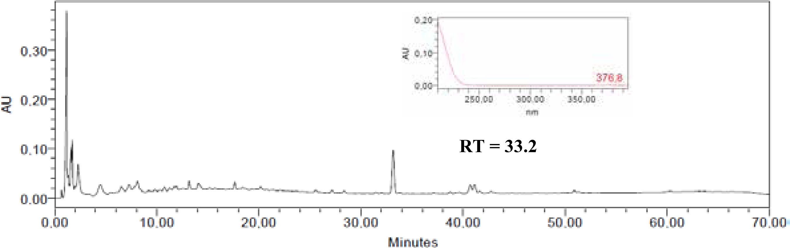
XM-1 is responsible for the peak at Rt 33.2 min in the HPLC-DAD profile of EEXMS (FIGURE 2) and its UV spectrum registered online shows only a terminal absorption curve that is coherent with the structure of arjunic acid. Importantly, EEXMS exhibited moderate activity against VACV (EC50 36.4 ± 3.7 mg/ml, IS = 13.7) but was inactive against DENV-2. This result is an indication of the low content of XM-1 in EEXMS. The AcOEt fraction was more potent than XM-1 disclosing an EC50 of 12.5 μg/mL and IS > 5 against DENV-2 that may be related to possible synergism with other compounds present in this fraction. The presence of other antiviral compounds in the AcOEt fraction can be inferred from its activity against VACV and HSV-1 while XM-1 has shown no effect against these viruses (TABLE 1).
Triterpenes as antivirals
There are few reports on the antiviral activity of triterpenes. Recently, Brandão and Collaborators (2013) reported the antiviral activity of ursolic acid against HSV-1 and dengue virus 2 (DENV-2) with EC50 values of 6.2 and 3.2 μg/mL, respectively.
Arjunic acid was firstly isolated from Terminalia arjuna (Roxb. ex DC.) Wight & Arn. and occurs in several other plant species (Verma et al., 2012). Triterpenes are a class of natural products that exhibit a broad spectrum of biological activities including anti-inflammatory, antimicrobial, anti-cancer, antimalarial and other activities (Lucetti et al., 2010; Manzano et al., 2013; Wu et al., 2013). Several triterpenes have shown antiviral activity (Cos et al., 2004; Jassim and Naji, 2003; Khan et al., 2005). The highly oxygenated triterpenes ganoderiol F and ganodermanontriol have been isolated from the fruits of Ganoderma lucidum and are active against HIV-1 (El-Mekkawy et al., 1998). Another triterpene with activity against HIV-1 is lancilactone C that inhibited the replication of this virus with an EC50 value of 1.4 μg/mL and a therapeutic index greater than 71.4 (Chen et al., 1999). Chiang and Collaborattors (2005) reported the broad spectrum antiviral activity of Ocimum basilicum L., the sweet basil of Indian and Chinese medicine, against diverse virus families. Aqueous and ethanolic extracts afforded ursolic acid that showed strong activity against HSV-1, adenovirus 8 (ADV-8), CVB1 and enterovirus 71 (EV71) with EC50 values of 6.6, 4.2, 0.4 and 0.5 μg/mL, respectively. The antiviral activity of ursolic acid against CVB1 and EV71 is evident during the infection process and the replication phase, indicating that ursolic acid can be a potential candidate against these RNA viruses, what deserves further investigation (Chiang et al. 2005).
Zhou and Collaborators (2010) reported two new antiviral triterpenes, shion-22-methoxy-20(21)-en-3-one and shion-22(30)-en-3,21-dione, isolated from the rhizomes of Aster tataricus. These compounds showed inhibitory effects against hepatitis B surface antigen (HBsAg) with EC50 values of 0.89 and 4.49 μg/mL, respectively. Shion-22-methoxy-20(21)-en-3-one inhibited hepatitis Be antigen (HBeAg) with EC50 value of 0.83 μg/mL, and shion-22(30)-en-3,21-dione showed inhibitory activity on hepatitis A (HAV) with an EC50 value of 11.2 μg/mL. Moronic acid which was isolated from Rhus javanica L. has shown activity against acyclovir-resistant, thymidine kinasedeficient and wild-type HSV-1 strains with EC50 of 1.6, 2.0 and 3.9 μg/mL, respectively. The betulonic acid isolated from this same species exhibited activity against wild-type HSV-1 (EC50 2.6 μg/mL). Oral administration of moronic acid to cutaneously HSV-1 infected mice significantly retarded skin lesions and/or prolonged the mean survival times of infected mice without toxicity. It can be considered a potential anti-HSV agent with a different mechanism of action than acyclovir, the main anti-herpes drug (Kurokawa et al, 1999).
The chloroform extract of Eriobotrya japonica (Thunb.) Lindl. contains some triterpene esters. Only 3-O-trans-caffeoyltormentic acid reduced rhinovirus infection. This compound was ineffective towards HIV-1 and sindbis virus replication (Tommasi et al., 1992).
Ursolic, oleanolic and betulinic acids and their derivatives occur frequently and sometimes abundantly in many plants and inhibit HIV-1 protease and the stability of the gp120/gp41 complex (Matthe'e; Wright and Konig, 1999; Labrosse, Treboute and Alizon, 2000; Cos et al., 2003; Yogeeswari and Sriram, 2005). Betulinic and oleanolic acids were isolated from Syzigium claviflorum (Roxb.) Wall. ex A.M. Cowan et Cowan. and exhibited anti-HSV and anti-HIV activity.
Betulinic acid is more active with an EC50 value of 1.4 μM (Ikeda et al, 2005; Chattopadhyay and Naik, 2007). Dihydrobetulinic acid has an EC50 of 0.9 μM, while the esterification at C-3 hydroxyl of those acids resulted in the more potent antiviral compound 3-O-(3,3'-dimethylsuccinyl) betulinic acid (DSB) with an EC50 < 3.5 x 10⁻⁴ pM. DSB can block a key step in the processing of a viral core capsid protein (Kashiwada et al. 1996) and is very active against drug-resistant virus, effective in an animal model of HIV infection and suiTABLE for use in combination therapy and is under phase II clinical trial.
Pavlova and Collaborators (2003) reported antiviral properties of betulin, betulinic and betulonic acids in cell cultures infected with HSV-1, influenza FPV/Rostock and ECHO 6 viruses. All the evaluated triterpenes were active against HSV-1. Betulin, and especially betulinic acid, also suppressed ECHO 6 virus replication.
Oleanolic acid isolated from many plants, including Xanthoceras sorbifolium Bunge wood (Sapindaceae), inhibits herpes and HIV virus replication, but oxidation at the C-3 hydroxyl position resulted in 3-oxotirucalla-7,24-dien-21-oic acid with improved antiviral activity (EC50 0.0039 μg/mL) and also block HIV protease with an IC50 of 10 μg/ mL (Labrosse, Treboute and Alizon et al., 2000; Yogeeswari and Sriram, 2005). Ursolic acid isolated from Crataegus pinnatifida Bunge leaves showed potent action against HIV-1 protease activity at 100 μg/mL (Kashiwada et al., 1996). Maslinic acid isolated from Geum japonicum Thunb. can inhibit HIV-1 protease at EC50 17.9 μg/mL while moronic acid extracted from Myrceugenia euosma (O. Berg) D. Legrand showed significant anti-HIV activity with therapeutic index greater than 186 (Ito et al. 2001; Xu et al., 1996). The protostanes, garcisaterpenes A and C, isolated from Garcinia speciosa Wall., showed significant inhibitory activities against HIV-1 RTase and in the syncytium formation assay, while a secocycloartene triterpenoid, nigronoic acid, from Schisandra sphaerandra Stapf, inhibits the RTase of both HIV-1 and HIV-2 (Rukachaisirikul et al. 2003; Sun et al., 1996).
Conclusion
The presently reported results identified the ethanol extract of X. myrianthum stems as a source of anti-dengue and anti-vaccinia virus compounds. Finally, our findings are in line with the traditional use of Bignoniaceae species as antiviral agents in different South American countries. To the best of our knowledge, the occurrence of arjunic acid in Bignoniaceae and the anti-DENV-2 activity are reported here for the first time.
Acknowledgements
This work was supported by funds from FAPEMIG - Fundação de Amparo à Pesquisa do Estado de Minas Gerais (Brazil) and CNPq - Conselho Nacional de Desenvolvimento Científico e Tecnológico.
References
Assis, F.L.; Borges, I.A.; Mesquita, V.S.; Ferreira, P.C.; Trindade, G.S.; Kroon, E.G.; Abrahão, J.S. 2013 - Vaccinia Virus in Household Environment during Bovine Vaccinia Outbreak, Brazil. Emerging Infectious Diseases, v.19, p.2045 - 2047.
Barbosa, W.L.R.; Pinto, L.N.; Quignard, E.; Vieira, J.M.V.; Silva Jr., J.O.C.; Albuquerque, S. 2008 - Arrabidaea chica (HBK) Verlot: phytochemical approach, antifungal and trypanocidal activities. Brazilian Journal of Pharmacognosy, v.18, p.544-548.
Betancur-Galvis, L.A.; Saez, J.; Granados, H.; Salazar, A.; Ossa, J.E. 1999 - Antitumor and Antiviral Activity of Colombian Medicinal Plant Extracts. Memórias do Instituto Oswaldo Cruz, v. 94, p. 531-535.
Brandão, G.C.; Kroon, E.G.; Santos, J.R.; Oliveira, A.B. 2010a - Antiviral activities of plants occurring in the state of Minas Gerais, Brazil: Part 2. Screening Bignoniaceae species. Brazilian Journal of Pharmacognosy, v.20, p. 742-750.
Brandão, G.C.; Kroon, E.G.; Santos, J.R.; Stehmann, J.R.; Lombardi, J.A.; Oliveira, A.B. 2010b - Antiviral activity of Bignoniaceae species occurring in the State of Minas Gerais (Brazil): part 1. Letters in Applied Microbiology, 51, 469-476.
Brandão, G.C.; Kroon, E.G.; Souza, D.E.R.; Souza Filho, J.D.; Oliveira, A.B. 2013 - Chemistry and Antiviral Activity of Arrabidaea pulchra (Bignoniaceae). Molecules, v.18, p.9919-9932.
Chattopadhyay, D.; Naik T.N. 2007 - Antivirals of Ethnomedicinal Origin: Structure-activity Relationship and Scope. Mini Reviews in Medicinal Chemistry, v.7, p.275-301.
Chen D.; Zhang, S.; Wang, H.; Zhang, S.; Sun, Q.; Cosentino, L.; Lee, K. 1999 - Novel anti-HIV lancilactone C and related triterpenes from Kadsura lancilimba. Journal of Natural Products, v.62, p.94-97.
Chiang, L.C.; Leng, L.T.; Cheng, P.W.; Chiang, W.; Lin, C.C. 2005 - Antiviral activities of extracts and selected pure constituents of Ocimum basilicum. Clinical and Experimental Pharmacology and Physiology, v.32, p.811-816.
Cos, P.; Maes, L.; Vanden Berghe, D.; Hermans, N.; Pieters, L.; Vlietinck, A.J. 2004 - Plant substances as anti-HIV agents selected according to their putative mechanism of action Journal of Natural Products, v. 67, p.284-293.
Cos, P.; Vanden Berghe, D.; De Bruyne, T.; Vlietinck, A.J. 2003 - Plant substances as antiviral agents: an update (1997-2001). Current Organic Chemistry, v.7, p.1163-1180.
Delgado, M.C.C.; Da Silva, M.S.; Fot, R.B. 1984 - 3p-hydroxy-21p-e-cinnamoyloxyolean-12-en-28-oic acid, a triterpenoid from Enterolobium contorstisiliquum. Phytochemistry, v.23, p.2289-2292.
Eldeen, I.M.; Van Heerden, F.R.; Van Staden, J. 2008 - Isolation and biological activities of termilignan B and arjunic acid from Terminalia sericea roots. Planta Médica, v.74, p.411-413.
El-Mekkawy, S.; Meselhy, M.R.; Nakamura, N.; Tezuka, Y; Hattori, M.; Kakiuchi, N.; Shimotohno, K.; Kawahata, T.; Otake, T 1998 - Anti-HIV-1 and anti-HIV-1-protease substances from Ganoderma lucidum. Phytochemistry, v.49, p.1651-1657.
Gentry, A.H. 1992 - A synopsis of Bignoniaceae ethnobotany and economic botany. Annals of the Missouri Botanical Garden, v.79, p.53-64.
Hussain, H.; Krohn, K.; Ahmad, V.U.; Miana, G.A.; Green, I.R. 2007 - Lapachol: an overview. Arkvoc, p.145-171.
Ikeda, T.; Yokomizo, K.; Okawa, M.; Tsuchihashi, R.; Kinjo, J.; Nohara, T.; Uyeda, M. 2005 - Anti-herpes virus type 1 activity of oleanane-type triterpenoids. Biological and Pharmaceutical Bulletin., v.28, p.1779-1781.
Ito, J.; Chang, F.R.; Wang, H.K.; Park, YK.; Ikegaki, M.; Kilgore, N.; Lee, K.H. 2001 - Anti-AIDS Agents. 48.1 anti-HIV activity of moronic acid derivatives and the new melliferone-related triterpenoid isolated from Brazilian propolis Journal of Natural Products, 64, 1278-1281.
Jassim, S.A.A.; Naji, M.A. 2003 - Novel antiviral agents: a medicinal plant perspective. Journal of Applied Microbiology, v.95, p.412-427.
Kashiwada, Y.; Hashimoto, F.; Cosentino, L.M.; Chen, C.H.; Garrett, P.E.; Lee, K.H. 1996 - Betulinic acid and dihydrobetulinic acid derivatives as potent anti-hiv agents. Journal of Medicinal Chemistry, v.39, p.1016-1017.
Kernan, M.R.; Amarquaye, A.; Chen, J.L.; Chan, J.; Sesin, D.F.; Parkinson, N.; Ye, Z.; Barrett, M.; Bales, C.; Stoddart, C.A.; Sloan, B.; Blanc, P.; Limbach, C.; Mrisho, S.; Rozhon, E.J. 1998 - Antiviral phenylpropanoid glycosides from the medicinal plant Markhamia lutea. Journal of Natural Products, v.61, p.564-570.
Khan, M.T.H.; Ather, A.; Thompson, K.D.; Gambari, R. 2005 - Extracts and molecules from medicinal plants against herpes simplex viruses. Antiviral Research, v.67, p.107-119.
Kumar, P.A.; Das, S.K. 1996 - A colorimetric assay to evaluate the cytotoxic activity of the intestinal intraepithelial lymphocytes of chickens. Veterinary Research Communications, v.20, p.513-518.
Kurokawa, M.; Basnet, P.; Ohsugi, M.; Hozumi, T.; Kadota, S;. Namba, T.; Kawana, T.; Shiraki, K. 1999 - Anti-herpes simplex virus activity of moronic acid purified from Rhus javanica in vitro and in vivo. The Journal of Pharmacology and Experimental Therapeutics, v.289, p.72-78.
Labrosse, B.; Treboute, C.; Alizon, M. 2000 - Sensitivity to a nonpeptidic compound (RPR103611) blocking human immunodeficiency virus type 1 Envmediated fusion depends on sequence and accessibility of the gp41 loop region. Journal of Virology, v.74, p.2142.
Lima, C.S.A.; Amorim, E.L.C.; Sena, K.X.F.R.; Chiappeta, A.A.; Nunes, X.P.; Agra, M.F.; Cunha, E.V.L.; Silva, M.S.; Barbosa-Filho, J.M. 2003 - Antimicrobial activity of a mixture of two isomeric phenylpropanoid glycosides from Arrabidaea harleyi A.H. Gentry (Bignoniaceae). Brazilian Journal of Pharmaceutical Sciences, v.39, p.77-81.
Lohmann, L.G. 2006 - Untangling the phylogeny of neotropical lianas (Bignonieae, Bignoniaceae). American Journal of Botany, v.93, p.304-318.
Lohmann, L.G. 2014 - Bignoniaceae in Lista de Espécies da Flora do Brasil. Jardim Botânico do Rio de Janeiro. Disponível em: <http://floradobrasil.jbrj.gov.br/jabot/fíoradobrasil /FB112305>. Acesso em: 01 Mar. 2014.
Lucetti, D.L.; Lucetti, E.C.P.; Bandeira, M.A.M.; Veras, H.N.H.; Silva, A.H.; Leal, L.K.A.M.; Lopes, A.A.; Alves, A.C.C.; Gabriela S Silva, G.S.; Brito, G.A.; Viana, G.B. 2010 - Anti-inflammatory effects and possible mechanism of action of lupeol acetate isolated from Himatanthus drasticus (Mart.) Plumel. Journal of Inflammation 7:60 Disponível em: <http://www.journal-inflammation.com/content/7/1/60> Acesso em 03 Dez. 2014
Mahato, S.B.; Kundu, A.P 1994 - 13C NMR spectra of pentacyclic triterpenoids - a compilation and same saliente features. Phytochemistry, v.37, p.1517-1575.
Manzano, P.I.; Miranda, M.; Abreu-Payrol, J.; Silva, M.; Sterner, O.; Peralta, E.L. 2013 - Pentaciclyc triterpenoids with antimicrobial activity from the leaves of Vernonanthura patens (Asteraceae). Emirates Journal of Food and Agriculture, v.25, p.539-543.
Matthée, G.; Wright, A.D.; Konig, G.M. 1999 - HIV reverse transcriptase inhibitors of natural origin. Planta Médica, v.65, p.493-506.
Oberste, M.S.; Blair, E.P.G.; Nix, A.W.; Ksiazek, T.G.; Comer, J.A.; Rollin, P.; Goldsmith, C.S.; Olson, J.; Kochel T.J. 2009 - Human febrile illness caused by Encephalomyocarditis Virus infection, Peru. Emerging Infectious Diseases, v.5, p.640-646.
Oliveira, A.B.; Raslan, D.S.; Miraglia, M.C.M.; Mesquita, A.A.L.; Zani, C.L.; Ferreira, D.T.; Maia, J.G.S. 1990 - Estrutura química e atividade biológica de naftoquinonas de Bignoniáceas brasileiras. Química Nova, v.13, p.302-307.
Pauletti, P.M.; Bolzani, V.S.; Young, M.C.M. 2003. Constituintes químicos de Arrabidaea samydoides (Bignoniaceae). Química Nova, v.26, p.641-643.
Pauletti, P.M.; Gamboa, I.C.; Silva, D.H.S.; Young, M.C.M.; Tomazela, D.M.; Eberlin, M.N.; Bolzani, VS. 2003 - New antioxidant C-Glucosylxanthones from the stems of Arrabidaea samydoides. Journal of Natural Products, v. 66, p. 1384-1387.
Pavlova, N.I.; Savinova, O.V.; Nikolaeva, S.N.; Boreko, E.I.; Flekhter, O.B. 2003 - Antiviral activity of betulin, betulinic and betulonic acids against some enveloped and non-enveloped viruses. Fitoterapia, v.74, p.489-492.
Pinto, A.V.; Pinto, M.C.F.R.; Lagrota, M.H.; Wigg, M.D.; Aguiar, A.N.S. 1987 - Antiviral activity of naphthoquinones: I. Lapachol derivatives against enteroviruses. Revista latinoamericana de microbiologia, v.29, p.15-20.
Rodriguez, D.J.; Chulia, J.; Simões C.M.O.; Amoros, M.; Mariotte, A.M.; Girre, L. 1990 - Search for in vitro antiviral activity of a new isoflavonic glycoside from Ulex europaeus. Planta Medica, v.56, p.59-62.
Rukachaisirikul, V.; Pailee, P.; Hiranrat, A.; Tuchinda, P.; Yoosook, C.; Kasisit, J.; Taylor, W.C.; Reutrakul, V. 2003 - Anti-HIV-1 protostane triterpenes and digeranylbenzophenone from trunk bark and stems of Garcinia speciosa. Planta Médica, v.69, p.1141-1146.
Silverstein, R.M.; Webster, F.X. (ed.) 2000 - Identificação espectrométrica de compostos orgânicos. Guanabara Koogan. Rio de Janeiro.
Sun, H.D.; Qiu, S.X.; Lin, L.Z.; Wang, Z.Y; Lin, Z.W.; Pengsuparp, T.; Pezzuto, J.M.; Fong, H.H.S.; Cordell, G.A.; Farnsworth, N.R. 1996 - Nigranoic acid, a triterpenoid from Schisandra sphaerandra that inhibits HIV-1 reverse transcriptase. Journal of Natural Products, v.59, p.525-527.
Teixeira M.T. 2012 - Few characteristics of dengue’s fever epidemiology in Brazil. Revista do Instituto de Medicina Tropical de São Paulo, v. 54(Suppl. 18), p.S1-S4.
The Plant List A working list of all plant species. 2014. http://www.theplantlist.org/tpl1.1/search?q=Xylophragma. Acesso 23/03/14.
Tommasi, N.; Simone, F.; Pizza, C.; Mahmood, N.; Moore, P.S.; Conti, C.; Orsi, N.; Stein, M.L. J. 1992 - Constituents of Eriobotrya japonica. A study of their antiviral properties. Journal of Natural Products, v.55, p.1067-1073.
Verma, S.C.; Jain, C.L.; Padhi, M.M.; Devalla, R.B. 2012 - Microwave extraction and rapid isolation of arjunic acid from Terminalia arjuna (Roxb. ex DC.) stem bark and quantification of arjunic acid and arjunolic acid using HPLC-PDA technique. Journal of Separation Science, v.35, p.1627-33.
Vlietinck, A.J.; Van Hoof, L.; Totte, J.; Lasure, A.; Vanden-Berghe, D.; Rwangabo, P.C.; Mvukiyumwami, J. 1995 - Screening of hundred Rwandese medicinal plants for antimicrobial and antiviral properties. Journal of Ethnopharmacology, v.46, p. 31-47.
Wagner, H.; Bladt, S.; Zgainsky, E.M. (ed.) 1984 - Plant drug analysis: a thin layer chromatography atlas. Springer. Berlin.
Whitley, R.J.; Roizman, B. 2001 - Herpes simplex virus infections. The Lancet, v.357; p.1513-1518.
Wu, G.S.; Guo, J.J.; Bao J.L.; Li, X.W.; Chen, X.P.; Lu, J.J.; Wang, Y.T 2013 - Anti-cancer properties of triterpenoids isolated from Ganoderma lucidum - a review. Expert Opinion on Investigational Drugs, v.22, p.981-992.
Yogeeswari, P.; Sriram, D. 2005 - Betulinic acid and its derivatives: a review on their biological properties Current Medicinal Chemistry, v.12, p.657-666.
Xu, H.X.; Zeng, F.Q.; Wan, M.; Sim, K.Y. 1996 - AntiHIV Triterpene acids from Geum japonicum. Journal of Natural Products, v. 59, p. 643-645.
Zhou, W.B.; Tao, J.T.; Xu, H.M.; Chen, K.L.; Zeng, G.Z.; Ji, C.J.; Zhang, Y.M.; Tan, N.H. 2010 - Three new antiviral triterpenes from Aster tataricus. Verlag der Zeitschrift für Naturforschung, v 65b, p.1393-1396.